
INTRODUCTION
By. Dr. Robert L. Bard
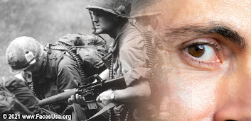
High risk professions like law enforcement, military service, healthcare and emergency response are known to have exposure to some of the most extreme levels of trauma - both physically and psychologically. They range in effects from manageable symptoms to crippling disorders. Over time, most people overcome disturbing or traumatic experiences and continue to work and live their lives. But others who get affected by traumatic experiences may trigger a reaction that can last for months or even years. This is called Post-traumatic Stress Disorder, or PTSD. Proportionately, studies have shown a lower percentage of retirees from such challenging careers acquire PTSD (from 15-20%) while an estimated 30-40% who suffer from PTSD associated symptoms go undetected or do not register as full cases. A larger percentage ‘on the job’ might be able to maintain the expected work standards throughout their career and even make it to retirement without visible signs. But “POST traumatic recall” leading to fully blown PTSD occurs when repeated exposure to trauma compounds on the tolerance capacity that eventually, one’s coping ability collapses. The individual may feel stages of grief, depression, anxiety, guilt or anger from uncontrollable issues like recurring flashbacks and nightmares.
National programs are now being explored to provide continued improvements in resources and access to mental health solutions. Additional projects are in place to offer advocacy and outreach to the rescue community to reduce and eliminate stigma, facilitating optimal ease and support to ASK FOR HELP.
PART 1: 2021 PROGRAM BLUEPRINT
By: Jessica Connell, LCSW, CPC, CEC
Until recently, I find that the vast majority of government-backed mental health programs were looked down upon in service communities like rescue workers and law enforcement. Upon interviews and sessions with retired firefighters for our EMDR review, I have identified the resistance from common concerns about being chastized socially or financially- from questions like "how does my mental health report affect my LIFE INSURANCE?... or PENSION?.. or my CURRENT JOB?"
Strategically, resolving this concern is crucial. As part of our focus to de-stigmatize GETTING HELP- we must first provide assurance and the feeling of safety in ASKING FOR HELP. This has become the central basis for the program we are shaping here.
THE SELECTION PROCESS
The engagement of the patient is everything. The wrong start can lead to disaster. The F.A.C.E.S. Network and I established an exploratory effort to assess and re-engineer a plan for a more effective support system and a public awareness program - making it more comfortable for any responder to ask for help. This next generation concept is with the full support of first responders health advocates and recognized supporters from the medical and mental health communities.
One key element in this plan that we are already seeing take place is the privacy and anonymity component. Upon exploring the ideal blueprint of an effective mental health program for active and retired rescue workers, my research had led me to what was once an experimental grant-funded national project with the Federal Park Ranger Service. A public resource was offered to this community to attend ANONYMOUSLY- outside the unit or official location(s). A designated therapist is available to the patient to privately undergo counseling, coaching and other modalities of therapy where each service personnel feels that form of safety from stigma. The project works and is continued today in a similar model and is adapted on a state by state level, and the model is also picked up by other service agencies.
Day One: volunteers warmed up to me and we explored that WARMING UP process. We discussed the community objective (for the firefighter), the current lack of initiatives to explore mental health issues and the heavy exposure on "the job" of the varying forms of trauma. This was the ideal common ground that helped to flesh out the camaraderie or teamwork that we needed and that which therapy is based on. For this healing process to happen, we need to work together.
The therapist-patient connection is as unique as it is crucial. There's many different kinds therapists out there- and to try to find the right one for you, that's a good fit. DAY ONE is that getting to know each other, where being on the same page as well as exploring that syneregy is key. It's a real relationship where you can really go back and forth where the patient needs to find that safe space with the therapist so they can talk about anything that they want.
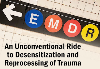
EMDR (Eye movement desensitization and reprocessing)
By introducing something innovative like EMDR puts the patient at the drivers seat as a segue to learning about the inroads of therapy because EMDR is so structured and less of the fear of "talk therapy" and opening up.
EMDR works on RECALL in a more structured way by targeting a specific event. It places the subject/patient in a somewhat dreamlike state (of calm) where we guide them through a specific event and try to untangle the trauma from the body- that trauma that is "trapped" in the nervous system. A good way to describe the experience is to create a form of fantasy version of this event and make it more safe for the person to review. Another good way to present the experience is much like a Virtual Reality simulation- but that YOU are doing the programming. EMDR unlocks the bottled trauma and the objective of the therapist (or guide) is to bring down the stigma incited by the patient about that event or issue, so we can safely and comfortable work on it.
In an array of interviews with active and retired first responders, the first step is to get them all comfortable with opening up. Some were more closed and guarded than others, but as first responders go, we are working with volunteers who are also health advocates and public caregivers, so the resistance is a lot less. Together we are partners to explore how to formulate an outreach and support program. These same volunteer patients will experience the benefits of mental health support to form the means of bringing it to those who need.
 Eye movement desensitization and reprocessing (EMDR) is a form of psychotherapy in which the person being treated is asked to recall distressing images; the therapist then directs the patient in one type of bilateral stimulation, such as side-to-side eye rapid movement or hand tapping. EMDR was developed in 1988. According to the 2013 World Health Organization (WHO): "This therapy [EMDR] is based on the idea that negative thoughts, feelings and behaviours are the result of unprocessed memories. The treatment involves standardized procedures that include focusing simultaneously on (a) spontaneous associations of traumatic images, thoughts, emotions and bodily sensations and (b) bilateral stimulation that is most commonly in the form of repeated eye movements." EMDR is included in several evidence-based guidelines for the treatment of post-traumatic stress disorder (PTSD), with varying levels of recommendation and evidence (very low to moderate per WHO stress guidelines). As of 2020, the American Psychological Association lists EMDR as an evidence-based treatment for PTSD but stresses that "the available evidence can be interpreted in several ways" and notes there is debate about the precise mechanism by which EMDR appears to relieve PTSD symptoms with some evidence EMDR may simply be a variety of exposure therapy. Even though EMDR is effective, critics call it a pseudoscience because only the desensitization component has scientific support. (Source: Wikipedia) |
By: Lt. Chris Conner/ Bedford Fire Dept (TX)


 ROBERT L. BARD, MD, PC, DABR, FASLMS - Advanced Imaging & Diagnostic Specialist
Having paved the way for the study of various cancers both clinically and academically, Dr. Robert Bard co-founded the 9/11 CancerScan program to bring additional diagnostic support to all first responders from Ground Zero. His main practice in midtown, NYC (Bard Diagnostic Imaging- www.CancerScan.com) uses the latest in digital Imaging technology has been also used to help guide biopsies and in many cases, even replicate much of the same reports of a clinical invasive biopsy. His most recent program is dedicated to the reporting of mental health diagnostic and innovative solutions including the use of modern neuromagnetic technologies and protocols in his MEDTECH REVIEWS program.
 JESSICA A. CONNELL, LMSW, CPC, CEC - Responders' Mental Health Program
As a therapist and coach, a lot of my work with clients is helping to manage symptoms of anxiety and panic- that which manifests in physical, often frightening and alarming ways. We can experience things like racing heartbeat, shortness of breath, numbness in arms and legs which can all make us feel like we are out control of our bodies and our surrounding world. When we have experienced a traumatic event in our lives, these feelings can be even more severe and heightened. The trauma and residually related fear is one that is very close to my heart and a reason I can provide empathy and understanding to clients that have been affected by the horrific day. When we work to process physical emotions that arise from trauma, the hope is that one day we can be less affected by it and live more presently to enjoy life’s fulfilling moments. I work with clients to slowly pull apart the physical emotions we experience from the thoughts that we are having and process them in a more self-aware and grounded way., visit her website- www.jagtheracoach.com


